Original article : L’apparato di Golgi: caratteristiche, struttura e funzioni by Silvia De Giorgi
Characteristics
One of the main activities carried out in the eukaryotic cell is surely the activity of protein synthesis. Protein production from the genome is the basis of cell life and its interactions with other cells. Indeed, the proteins constitute the organism and can be of different types: structural, functional, enzymatic, transport, reserve, defense.
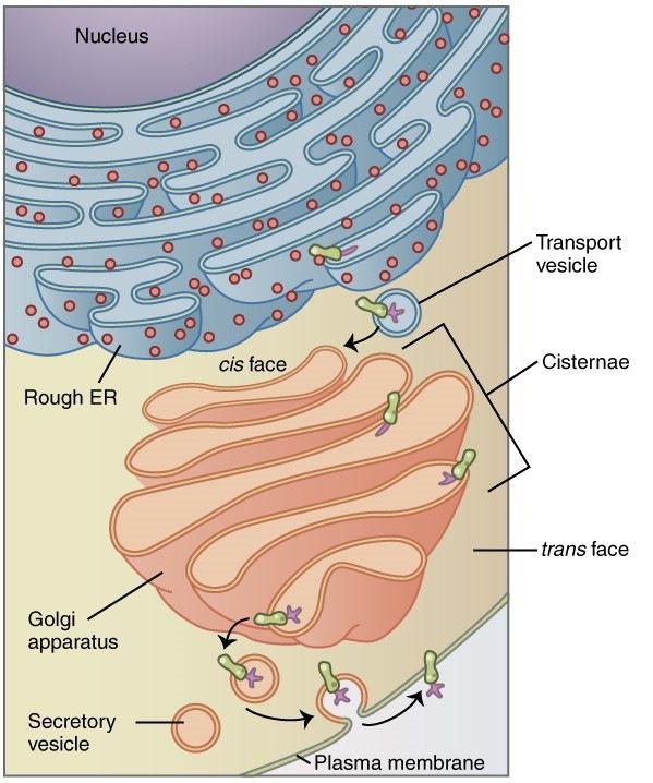
In the production process of a protein, there are several stages that lead to its maturation and final functional form. One of the organelles involved in these processes is the Golgi apparatus: here the neosynthesized proteins come in vesicles from the endoplasmic reticulum and are reworked and sorted to different cell compartments or to the plasma membrane.
It was the physician and histologist Camillo Golgi who first identified and described this organelle, already before the advent of the electron microscope, using specific histological stainings (Fig.1).

The Golgi apparatus is a real sorting center, which at the same time allows the maturation processes of proteins, thus dealing with post-translational modifications. The compartments of this structure are functionally differentiated, allowing proteins to be assigned to different intra and extracellular localizations.
Structure
The apparatus of Golgi (Fig. 2) is constituted by a pile of flattened vesicles and delimited by a membrane. These are called cisternae, and besides these are present a series of vesicles with a diameter of less than 100 nm, the transport vesicles, and other larger, called secretion vesicles.
One distinguishes a proximal face, directed towards the RER, also called cis-face, and a distal, or trans-face, of maturation. The cell faces the surface of the cell (Fig. 3). Usually, this apparatus is localized close to the nucleus, to be able to “receive” from the rough endoplasmic reticulum (RER), rich in ribosomes, the amino acid chains just synthesized, through vesicles.
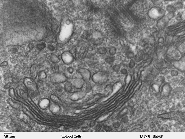
The number of cisternae generally varies from four to six, but there are cells with even a dozen of cisternae. It goes without saying that Golgi’s apparatus is much more complex and developed in cellular types engaged in intense protein activity.
Functions
Through gemmation and endocytosis events, the vesicles carrying the protein material, deriving from the RER, merge with the cisternae in cis of the Golgi apparatus. Along with it then, the proteins undergo various modifications until leaving the trans cisternae directed towards the various cellular components. This intense vesicular traffic and the transit through the cisternae are assisted by the cytoskeleton components. Sorting is possible thanks to various molecular components that identify the path of the vesicle. They can be directed to other cell organelles, to the cytoplasmic membrane, or to the outside of the cell.
Changes to proteins that occur at the level of the Golgi apparatus may be additions or even simplifications, depending on the fate of the protein. From the apparatus of Golgi originate therefore two families of molecules: oligosaccharides with high content of mannose and oligosaccharides complexes. In the case of the first category the chains do not meet with further additions in the apparatus; in the second case instead, three units of mannose are eliminated, but at the same time other trisaccharide units are added.
Depending on the destination of the protein, then, further, and different modifications can take place. In the case of lysosomal target proteins, for example, mannose residues are phosphorylated.
Luigi Gallucci
Bibliography
- Cell biology, Colombo R., Elm E. (link)
- Featured image: Wikimedia Commons